An Overview on Ankle Anatomy
The ankle is a sophisticated structure composed of two separate joints – the tibiotalar joint and the subtalar joint. Acting like a hinge, the unique ankle anatomy allows the joint to withstand 1.5 times the body’s weight when walking and up to 8 times the body’s weight when running. A complex system of bones, ligaments, tendons and muscles make up the ankle that provides the ability to walk, run and exercise. Injuries to these structures will often lead to ankle pain and loss of function. Santa Barbara, Goleta, Santa Maria and Ventura, California orthopedic ankle specialist, Dr. Jervis Yau is highly experienced at diagnosing and treating ankle injuries and conditions.
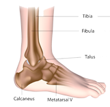
Anatomy of the Ankle
Ankle Bones and Joints
The true ankle joint consists of three bones – talus (ankle bone), tibia (shinbone) and fibula (lower leg bone). The top of the talus fits inside a socket formed by the lower portion of the tibia and fibula. The bottom of the talus sits on the calcaneus (heel bone). The talus plays an important role in ankle function by allowing the joint to move the foot up and down.
Beneath the true ankle joint is the subtalar joint, which is formed by the articulation of the talus and calcaneus. The subtalar joint allows for inversion and eversion of the foot (side to side).
Ankle Articular Cartilage
Articular cartilage covers the ends of each bone in the ankle and allows the bones to move pain-free and smoothly against each other. The cartilage helps the joint carry weight by providing shock absorption.
Ankle Ligaments, Muscles and Tendons
Three ligaments are involved in the lateral ligament complex and include the anterior talofibular ligament (ATFL), the calcaneofibular ligament (CFL) and the posterior talofibular ligament (PTFL). These three ligaments are located on the lateral (outside) portion of the ankle.
Three ligaments support the articulation of the fibula and tibia, known as the ankle syndesmosis. These ligaments include the anterior inferior tibiofibular ligament (AITFL), the posterior inferior tibiofibular ligament (PITFL) and the transverse ligament.
Important muscles and its associated tendons include the following. The gastrocnemius and soleus muscles connect to the calcaneus through the Achilles tendon allowing for plantarflexion and push off strength. The peroneal longus and brevis attach to the midfoot and allows for dorsiflexion and eversion. The tibialis anterior is the primary ankle dorsiflexor while the tibialis posterior allows for plantarflexion, inversion and arch support.
The ankle is in constant motion and thus, prone to overuse injuries and conditions in the active population. When the joint becomes damaged from injury, overuse, trauma or natural degeneration, patients may experience pain, instability, weakness and loss of mobility.
Dr. Yau commonly treats the following ankle conditions:
To learn more about ankle anatomy, or to diagnose an injury or condition causing your ankle pain, please contact the orthopedic office of Dr. Jervis Yau located in the Santa Barbara, Goleta, Santa Maria and Ventura, California communities.