An Overview on Elbow Anatomy
Located in the center of the arm, the elbow is one of the most widely used joints in the human body. The elbow joint is a relatively simple hinge joint composed of bones, ligaments, tendons, muscles and nerves. All of these structures work in unison to allow the arm to bend and move. This allows Santa Barbara, Goleta, Santa Maria and Ventura, California patients to engage in athletic activities. When one or more of these structures gets damaged from normal wear and tear or sports, patients may develop elbow pain and loss of function. Dr. Jervis Yau, orthopedic elbow specialist, specializes in elbow anatomy and the many injuries and conditions that affects the joint.
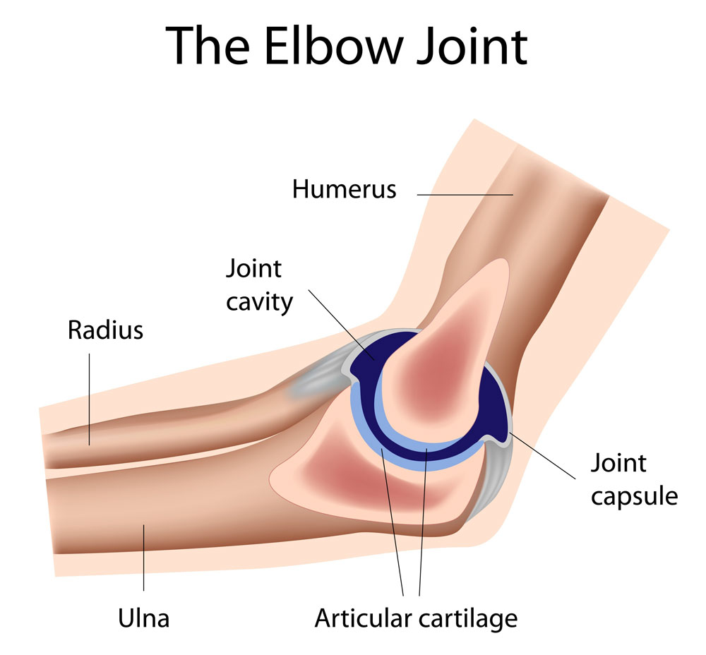
Anatomy of the Elbow
The shoulder, elbow and wrist work in unison to provide function to the arm. The elbow can flex and extend while the forearm can pronate and supinate (rotation) allowing for multiple degrees of motion.
The elbow joint consists of three bones:
- Humerus – The arm’s largest bone that connects to the shoulder to the forearm
- Radius – One of the two bones in the forearm that is located on the side of the thumb.
- Ulna – One of the two bones in the forearm that is located on the side of the pinky.
Tough, flexible ligaments allow for function and stabilizes the elbow to prevent injuries. The three main ligaments are:
- Ulnar collateral ligament (UCL) – Located on the elbow’s inner side to prevent valgus deformity
- Radial collateral ligament – Located on the joint’s outer side to prevent varus deformity
- Annular ligament – This ligament keeps the radial head in contact with the ulna during motion and rotation.
The remainder of elbow is composed of sophisticated structure of tendons, muscles and nerves, including:
- The flexors and extensors allow for movement of the wrist and fingers
- The biceps tendon allows the elbow to flex and the forearm to supinate
- The triceps tendon allows for elbow extension
- The median, ulnar and radial nerves provide motor function and sensation to the forearm, wrist and hand.
Elbow pain and dysfunction is commonly seen in athletes and non-athletes alike. Dr. Yau commonly treats the following causes of elbow pain:
- Distal biceps rupture
- Medial epicondylitis (Golfer’s elbow)
- Lateral epicondylitis (Tennis elbow)
- Ulnar collateral ligament injuries
- Cubital tunnel syndrome (Ulnar nerve entrapment)
For more resources on elbow anatomy or to determine the cause of your elbow pain, please contact the Santa Barbara, Goleta, Santa Maria and Ventura, California orthopedic office of elbow specialist Dr. Jervis Yau.