An Overview on Knee Anatomy
The knee is the largest joint in the human body and is easily injured because of its complex structure. The sophisticated anatomy of the knee joint allows it to flex, extend, twist from side to side and handle a significant amount of stress during athletic and everyday activities. Santa Barbara, Goleta, Santa Maria and Ventura, California orthopedic knee specialist, Dr. Jervis Yau specializes in diagnosing and treating various conditions that can cause pain and dysfunction of the knee.
Anatomy of the Knee
The knee joint is composed of bones, cartilage, ligaments and tendons. These structures work together to provide stability and fluidity in movement.
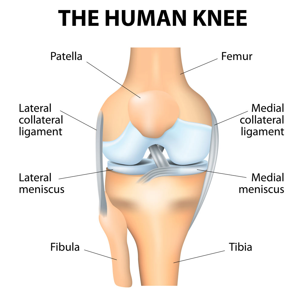
Bones meet to form the knee joint. These bones include the femur (thigh bone), tibia/fibula (shin bone) and patella (knee cap). Articular cartilage lines the ends of each bone to allow low friction gliding and force dispersion across the knee during movement and loading.
Another important form of cartilage that distributes load within the knee is the meniscus. The medial and lateral meniscus are two wedge-shaped pieces of cartilage that act as “shock absorbers” between the femur and tibia. Unlike smooth articular cartilage, meniscal cartilage is rubbery and tough to provide cushion and stabilization to the joint.
The bones are connected to each other through the joint capsule and four major ligaments that act as strong ropes to stabilize the joint. The cruciate ligaments (ACL and PCL) control the back and forth motion, while the collateral ligaments (MCL and LCL) control the side to side motion.
The four main knee ligaments are:
- Anterior cruciate ligament (ACL) – The ACL travels from the anterior tibia to the posterior femur and prevents the tibia from moving too far forward in relation to the femur.
- Posterior cruciate ligament (PCL) – The PCL travels from the posterior tibia to the anterior femur and wraps around the ACL, preventing the tibia from moving too far backwards in relation to the femur.
- Medial collateral ligament (MCL) – Consists of a superficial and a deep band that runs between the medial aspect of the tibia and femur to prevent the knee from collapsing in.
- Lateral collateral ligament (LCL) – The LCL connects the lateral femur to the fibular head and prevents the knee from collapsing out.
Tendons and muscles about the knee provide movement and function. Tendons connect muscles to bones and allow the knee to flex and extend. They also serve as secondary stabilizers for the joint during movement.
The complex structures of the knee can be injured when abnormal forces are applied through traumatic accidents, sports injury, overuse and natural wear and tear associated with aging.
Common knee injuries treated by Dr. Yau include:
- ACL injuries
- MCL injuries
- Multi-ligament knee injuries
- Meniscus tears
- Cartilage injuries
- Malalignment of the lower extremity
- Fractures
To learn more about knee anatomy, or for additional resources on common injuries of the knee joint, please contact Dr. Jervis Yau, orthopedic knee specialist serving the Santa Barbara, Goleta, Santa Maria and Ventura, California communities.