An Overview on Hip Anatomy
The hip is classified as a ball and socket joint that allows a wide range of motion for walking, running, squatting and performing numerous sports activities. The hip joint is composed of sophisticated structures including bones, ligaments, tendons and muscles. Santa Barbara, Goleta, Santa Maria and Ventura, California orthopedic hip surgeon, Dr. Jervis Yau specializes in treating injuries and conditions associated with hip pain and dysfunction.
Anatomy of the Hip
Hip Bones
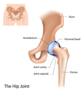
The hip is the largest ball and socket joint in the body and its complex structure allows patients to perform a variety of everyday activities. The hip joint is formed by the femur (thighbone) meeting the pelvis. The ball is the rounded end of the femur (femoral head) and the socket is a concave pocket in the pelvis’ lower side, otherwise known as the acetabulum. The femoral head is attached to the rest of the femur by a short portion called the femoral neck. Next to the femoral neck, a large bump located on the hip’s outer portion is a muscle attachment site, known as the greater trochanter.
Hip Articular Cartilage
Articular cartilage covers the ends of each bone in the hip and allows the bones to glide over each other in a fluid and pain-free motion. The smooth, white cartilage is lubricated by synovial fluid, which helps the hip joint withstand great forces associated with motion and loading during normal function and sports.
Hip Ligaments and Tendons
The hip joint is reinforced by a number of strong ligaments surrounding the joint and forming the joint capsule. These ligaments provide stabilizing support for the hip and are known as the pubofemoral, iliofemoral and ischiofemoral ligaments.
The tensor fascia lata and iliotibial band connects from the hip to the knee and provides support to the pelvis during gait.
Hip Muscles
A complex structure of muscles surrouding the hip joint allows it to bend, straighten and move with power and fluidity. The major hip muscles can be grouped according to its function.
Hip Abductors: Gluteus maximus, gluteus medius, gluteus minimus, tensor fascia lata
Hip Adductors: Adductor magnus, adductor longus, adductor brevis, pectineus, gracilis
Hip flexors: Iliopsoas (psoas major and minor, iliacus), rectus femoris, sartorious
Hip extensors: Gluteus maximus, biceps femoris, semimembranosus, semitendinosus
As an extremely mobile joint in the human body, the hip is in constant motion and prone to a number of injuries and conditions. When the hip joint becomes damaged from sports injury, overuse, trauma or aging, patients may experience pain, weakness, instability and loss of mobility.
Dr. Yau commonly treats these hip injuries and conditions:
- Snapping hip syndrome
- Psoas impingement/tendonitis
- Femoroacetabular impingement (FAI)
- Gluteus medius and minimus tears
- Proximal hamstring tears
- Articular cartilage injuries
- Trochanteric pain
To learn more about hip anatomy, or to diagnose an injury or condition associated with your hip joint, please contact the orthopedic office of Dr. Jervis Yau, hip surgeon located in the Santa Barbara, Goleta, Santa Maria and Ventura, California communities.
